Fluorescence Microscopes
Structured Illumination Microscopy – What Is It and How Does It Work?
Structured Illumination Microscopy (SIM) is a cutting-edge optical imaging technique that bridges the gap between conventional light microscopy and super-resolution methods. SIM enhances image resolution beyond the diffraction limit of light, allowing scientists to explore biological structures and processes with remarkable clarity. This article will delve into what structured illumination microscopy is, how it works, its benefits, and its applications in biological research.
What is Structured Illumination Microscopy?
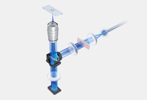
Example of SIM setup within an optical system
SIM is a super-resolution microscopy technique that improves spatial resolution by leveraging patterned illumination. Conventional light microscopes are limited by the diffraction of light, which caps its resolution at approximately 200 nm laterally and 500 nm axially. SIM bypasses this limitation by using interference patterns to encode high-resolution information that is then computationally reconstructed.
These interference patterns can be produced using a mechanical device with slits, laser projection, or other optical elements. The advantage of optically produced patterns over mechanical is that you can have more flexibility in the type of pattern that’s used – slit vs grid or pinhole – with the latter capable of providing better resolution on complex samples.
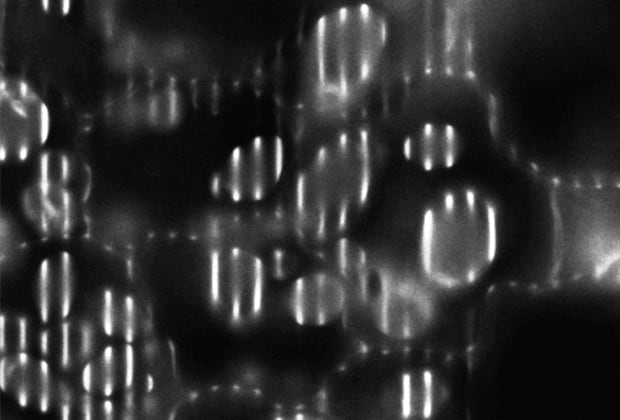
Slit projection
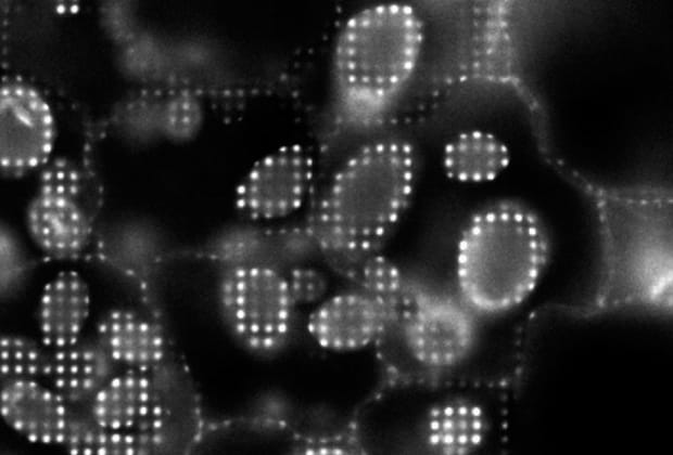
Grid/pinhole projection
This makes SIM ideal for imaging intricate biological structures, such as cytoskeletal filaments, organelles, and protein complexes, which are often smaller than the diffraction limit. Additionally, utilizing structured illumination within fluorescence microscopy can be critical to capture a clear image when working with thicker cells or tissues (organoids are a great example).
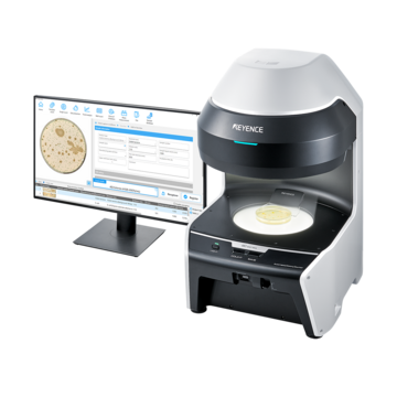
The BZ-X All-in-one Fluorescence Microscope from KEYENCE employs an optional electronic projection element to optically create slits or grid patterns. This allows users to capture high-resolution images without fluorescence blurring, even on thicker tissues. Contact us to learn more about this technology and how it can improve your research.
Contact us to learn more about how our advanced technology can help take your business to the next level.
Contact Us
How Does Structured Illumination Microscopy Work?

1. Patterned Illumination
The sample is illuminated with a known structured light pattern, often stripes or grids, which interact with the sample’s features to generate moiré fringes.
2. Image Acquisition
Multiple images are captured, each with a different phase or orientation of the structured pattern.
3. Image Reconstruction
Advanced algorithms decode the high-frequency spatial information embedded in the moiré fringes, reconstructing a super-resolved image. This step improves both lateral and axial resolution.
4. Multicolor and 3D Capabilities
SIM can also be adapted for multicolor imaging and three-dimensional (3D) reconstruction, enabling visualization of complex biological processes in real-time.
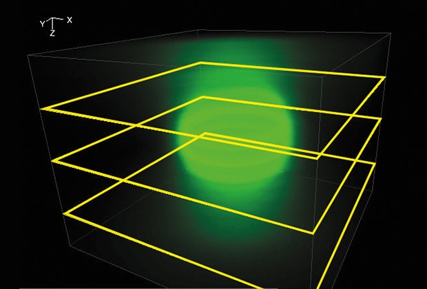
Epifluorescent 3D image
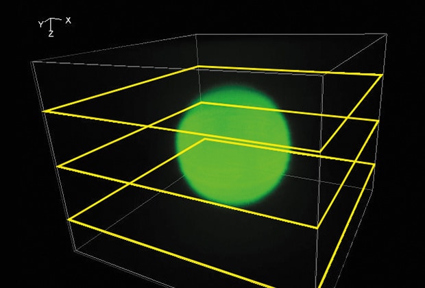
SIM 3D image
Discover more about this product.
Click here to book your demo.

Benefits and Advantages of Structured Illumination Microscopy
- Super-Resolution with Minimal Phototoxicity: SIM offers a significant improvement in resolution compared to conventional light microscopy without the high phototoxicity and photobleaching associated with techniques like Stimulated Emission Depletion (STED) or Single-Molecule Localization Microscopy (SMLM). This makes SIM particularly suited for live-cell imaging.
- Compatibility with Standard Fluorophores: Unlike other super-resolution techniques that require specialized dyes, SIM works with common fluorophores, making it accessible and cost-effective. This gives users access to the full fluorescence spectrum using a standard broad wavelength light source.
- High-Speed Imaging: SIM can capture super-resolved images relatively quickly, making it suitable for dynamic processes such as cell division, vesicle trafficking, or cytoskeletal remodeling.
- Ease of Use: Many modern SIM systems are user-friendly and compatible with standard microscopes, requiring only additional hardware and software.
- Multiscale Imaging: SIM enables researchers to study cellular events at both macro and micro levels, bridging the gap between conventional and electron microscopy.
We’re here to provide you with more details.
Reach out today!

Applications in Biological Research
Cellular and Subcellular Imaging
SIM is widely used to investigate cellular architecture, such as the organization of organelles like the endoplasmic reticulum, mitochondria, and Golgi apparatus.
Neuroscience
The technique has been pivotal in studying synaptic connections and neuronal dynamics, offering insights into brain function and diseases.
Developmental Biology
Researchers employ SIM to observe embryonic development and morphogenesis, uncovering the spatiotemporal coordination of cellular processes.
Pathogen-Host Interactions
SIM has advanced our understanding of how pathogens interact with host cells, aiding in the development of treatments for infectious diseases.
Structural Biology
High-resolution imaging of protein complexes and cytoskeletal components helps elucidate molecular mechanisms in health and disease.
Live-Cell Imaging
With its low phototoxicity, SIM is ideal for studying dynamic biological events such as intracellular transport, cell motility, and membrane dynamics.
Curious about our pricing?
Click here to find out more.

Structured Illumination Microscopy – Now You Know!
Structured Illumination Microscopy is a transformative tool in biological research, offering enhanced resolution, versatility, and live-cell imaging capabilities. Its ability to capture detailed images of cellular structures and processes has expanded our understanding of biology at a molecular level. As SIM technology continues to evolve, its applications are expected to grow, driving innovations in cell biology, neuroscience, and beyond.
KEYENCE’s BZ-X Automated Fluorescence Microscope functions like an automated fluorescence microscope system for routine epifluorescence imaging; however, it also has the modularity to optically section and reconstruct complex samples using structured illumination. This makes it the ideal system for those that are looking for a workhorse microscope which can also bridge the gap between epifluorescence and confocal microscopes.
Learn more about the BZ-X Automated Fluorescence Microscope by requesting an onsite or virtual demo or by downloading the product brochure.
Contact us to learn more about how our advanced technology can help take your business to the next level.
Contact Us

