Fluorescence Microscopes
Epifluorescence Microscopes vs. Confocal Microscopes: Which Is Right for You?
Microscopy is an essential tool in scientific research, and two of the primary fluorescence-based techniques are epifluorescence microscopy and confocal microscopy. Researchers widely use both for visualizing biological samples, but each has distinct advantages and limitations. This article will explore the main differences between epifluorescence and confocal microscopes, and help you decide which is best for your research needs.
What Is Epifluorescence Microscopy?
Epifluorescence, or widefield, microscopy is a type of light microscopy that uses fluorescence to illuminate specimens. It typically employs a mercury, xenon, or LED light source and filters to excite fluorophores within a sample, producing bright, high-contrast images against a dark background.
Advantages of Epifluorescence Microscopy
- Speed and Simplicity: Epifluorescence microscopes are user-friendly and provide quick imaging, making them ideal for routine diagnostics and live-cell imaging.
- Cost-Effective: These microscopes are generally less expensive than confocal microscopes, both in initial purchase and maintenance.
- Wide Field of View: They allow imaging of larger areas, suitable for applications like tissue section analysis.
Limitations of Epifluorescence Microscopy
- Out-of-Focus Light: The entire sample is illuminated, which means fluorescent signals from outside the focal plane can reduce image clarity.
- Limited Resolution: While sufficient for many applications, the resolution is lower compared to confocal microscopy.
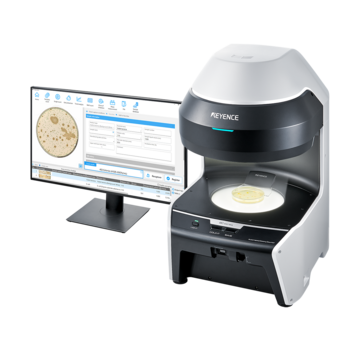
Our BZ-X All-in-One Fluorescence Microscope provides all the functionality of an epifluorescence microscope without the limitations. Contact us to learn more!
Contact us to learn more about how our advanced technology can help take your business to the next level.
Contact Us
What Is Confocal Microscopy?
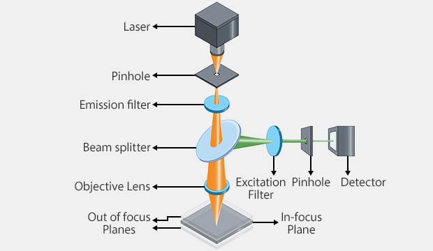
Basic operating principle of a confocal microscope
Confocal microscopy builds upon the principles of epifluorescence by introducing a pinhole aperture that eliminates out-of-focus light. This system scans samples point by point and reconstructs the image digitally, providing exceptional resolution and depth.
Advantages of Confocal Microscopy
- High Resolution and Contrast: By removing out-of-focus light, confocal microscopes deliver sharper and more detailed images.
- 3D Imaging: Confocal systems enable optical sectioning, allowing for three-dimensional reconstruction of samples.
- Customizable Imaging: Users can control parameters like pinhole size and laser excitation wavelengths for specific imaging needs.
Our BZ-X All-in-One Fluorescence Microscope is able to perform optical sectioning using structured illumination (optional), or SIM, in order to capture high-resolution images even on thicker samples. This technique also removes out-of-focus light to generate better spatial resolution without many of the drawbacks of confocal microscopes.
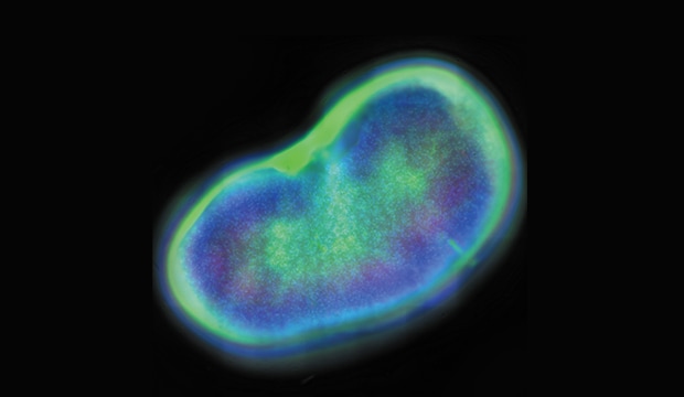
Conventional epifluorescence image
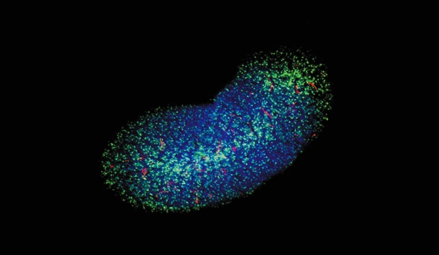
Structured illumination image (BZ-X)
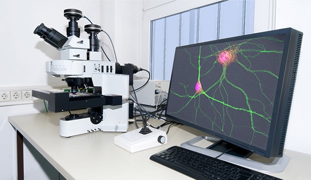
Example of confocal microscope on lab bench
Limitations of Confocal Microscopy
- High Cost: Confocal microscopes are significantly more expensive, both in terms of equipment and operational costs.
- Slower Imaging: The scanning process can be time-consuming, which may not be suitable for dynamic, fast-moving samples.
- Steep Learning Curve: Due to the complexity and customizations of these microscopes, users need to be thoroughly trained on their operation in order to capitalize on their functionality.
We’re here to provide you with more details.
Reach out today!

Epifluorescence vs. Confocal Microscopy: A Feature Comparison
| Feature | Epifluorescence | Confocal |
|---|---|---|
|
Resolution
|
Epifluorescence
Moderate
|
Confocal
High
|
|
Field of View
|
Epifluorescence
Wide
|
Confocal
Narrow (localized to focal planes)
|
|
3D Imaging
|
Epifluorescence
Available but limited
|
Confocal
Available
|
|
Cost
|
Epifluorescence
Lower
|
Confocal
Higher
|
|
Speed
|
Epifluorescence
Faster for basic imaging
|
Confocal
Slower due to scanning
|
|
Phototoxicity
|
Epifluorescence
Low
|
Confocal
High (due to laser)
|
Curious about our pricing?
Click here to find out more.

Applications in Research and Diagnostics
- Epifluorescence Microscopy: Commonly used in clinical and basic research settings for routine analysis, bacterial colony counting, traceability, and cell staining techniques. It's a go-to method for researchers who need fast, broad observations.
- Confocal Microscopy: Ideal for neuroscience, developmental biology, and cancer research, where fine details and three-dimensional structures are critical.
Discover more about this product.
Click here to book your demo.

Which Microscope Should You Choose?
Understanding the differences between epifluorescence microscopes and confocal microscopes is essential for selecting the right tool for your research or diagnostic needs. While epifluorescence microscopy excels in simplicity and cost, confocal microscopy offers unmatched image clarity and depth for complex analyses.
By leveraging the strengths of each, researchers can advance fields ranging from microbiology to molecular biology, ensuring accurate and efficient results.
Although choosing the right system for your research needs, both now and in the future, can be quite nuanced, the choice between an epifluorescence microscope and confocal microscope can generally be boiled down to:
- Choose epifluorescence microscopy if you need quick, cost-effective imaging of your samples.
- Opt for confocal microscopy when high resolution, depth, and precision are priorities.
The best recommendation is to make sure that you are thoroughly evaluating the options that can solve the majority of your requirements by bringing the systems into your lab and testing them on your own samples. This will give you firsthand experience on the capabilities of each microscope and allow you and any other lab members to understand how it would be used to enhance their research.
KEYENCE’s BZ-X Automated Fluorescence Microscope combines many of the capabilities and benefits of both epifluorescence and confocal microscopes into a single, modular platform. At its core, users can quickly and easily capture high-resolution, publication-quality images over wide areas in fluorescence, brightfield, and phase-contrast imaging methods.
This All-in-One Fluorescence Microscope can be used for routine checks or expanded to accommodate advanced techniques including live-cell incubation and time-lapse imaging, optical sectioning similar to a confocal, low-volume slide scanning, and image cytometry. With an integrated darkroom, users can install this system on any benchtop while taking up minimal laboratory space.
Discover why the BZ-X is the ideal choice for your imaging and high-resolution analysis needs. Download the brochure or request a demo in your lab.
Contact us to learn more about how our advanced technology can help take your business to the next level.
Contact Us

