Digital Microscopes
Optical Microscopes
Key Takeaways
- Optical microscopes use light and glass lenses to magnify samples, making them a foundational tool for visual analysis and imaging.
- Optical light microscopes are widely used for creating detailed micrographs in scientific, industrial, and research applications.
- Understanding how optical microscopes work helps users better evaluate image quality, magnification, and system capabilities.
- Comparing optical and digital microscopes is important for selecting the right tool based on workflow, analysis needs, and imaging requirements.

This article explores the fundamentals of optical microscopy, from basic lens principles to modern applications. You'll learn how light-based imaging systems create magnified views, when to choose optical versus digital platforms, and what factors determine image quality in different microscopy setups.
An Introduction to Optical Microscopes
What Is an Optical Microscope?
Optical microscopes are the most common type of microscope. Optical microscope is the general term used for microscopes that use visual light and glass lenses to perform magnified observation of objects.
They are also referred to as biological microscopes because they have been historically used to observe organisms such as microorganisms and the cells of plants and animals.
An optical microscope represents the most accessible form of high-magnification imaging. These instruments have served laboratories for centuries, evolving from simple single-lens designs to sophisticated multi-lens systems. Unlike specialized tools such as electron microscopes, which require vacuum chambers and extensive sample preparation, optical microscopes operate under standard room conditions with minimal specimen preparation.
This technology remains relevant because it strikes a balance between capability and practicality. Optical microscopes are used in research facilities to evaluate samples before moving to more specialized equipment. Quality control departments use them for routine inspections, where speed is more important than atomic-level imaging and data resolution. Educational institutions use optical microscopes to teach basic microscopy principles because students can view specimens through eyepieces, developing intuition regarding magnification and sample preparation in ways that digital displays cannot.
History of Optical Microscopes
Optical microscopes have a long history starting in 1590 when Hans Jansen and his son, Zacharias Jansen, created the first version using lenses in a tube, which allowed them to magnify and see small objects. In the latter half of the 17th century, Robert Hooke created the compound microscope by combining two lenses: an objective lens and an eyepiece, and used it to observe the structure of cork. Because the structure appeared to be a collection of small rooms like a beehive, he named these rooms cells. This led to the use of the word cell in biology. Around the same time, Antonie Philips van Leeuwenhoek created the single-lensed microscope (a microscope with one lens) and discovered microorganisms and sperm - the start of microbiology.
Modern optical microscopes are generally compound microscopes that combine an objective lens and an eyepiece. With typical optical microscopes, the light source is located below the sample, and it is observed by using the objective lens to magnify the light that is transmitted through the sample. Therefore, it is not possible to observe objects that do not transmit light. To observe such samples, it is necessary to cut them into thin slices and secure them on glass slides or similar objects. For samples that cannot be processed into thin slices and don’t transmit light, stereoscopic microscopes must be used.
Stereoscopic microscopes are optical microscopes that project light down onto the sample. The reflected light is then magnified by the objective lens for observation. They have two eyepieces, allowing for 3D observation that is the same as viewing the sample with the naked eye. Stereoscopic microscopes are used for relatively low-magnification observation.
Optical Light Microscopes
An optical light microscope achieves magnification through a two-stage lens system. Light passes through the specimen and enters the objective lens, which creates a magnified image. The eyepiece then magnifies this image further, producing the final view seen by the observer. Total magnification equals the objective power multiplied by the eyepiece power, so a 40x objective paired with a 10x eyepiece delivers 400x magnification.
What you see at high magnification depends on the quality of your lenses. Simple glass components used in less expensive systems cause chromatic aberration, which blurs small details and causes different colors to focus at various distances. To address these distortions, better objectives use several lens elements with unique coatings. Numerical aperture is also important.
The amount of light that the lens collects is indicated by this specification. Immersion oil between the lens and specimen is necessary for optimal performance. However, higher numerical aperture values between 0.95 and around 1.4 capture more light and produce sharper photos, without the need for immersion oil.
How Optical Microscopes Work
Understanding the light path clarifies what optical microscopes can and cannot do. Illumination from below passes through a condenser that focuses light onto the specimen. Transparent or semi-transparent samples allow light through, which the objective lens collects and magnifies. The physical wavelength of visible light sets an absolute resolution limit of around 200 nanometers because features closer together than that distance blur into a single point, regardless of magnification level.
This explains why increasing magnification beyond certain limits produces larger but not clearer images. A 1000x view might look impressive, but it reveals nothing more than a 400x view if the optics have reached their resolution threshold.
What is an optical microscope's practical limit? The magnification you need depends on what you're examining. Plant tissue work typically stays between 40x and 400x. Cell structure requires a higher magnification of around 400x to 600x. Viewing bacteria pushes the limits to approximately 1000x, which requires oil immersion lenses for the resolution needed at that level. These high-magnification applications demand careful technique and pristine optics to deliver useful results.
Applications of Optical Microscopes
Optical microscopes can be used for a wide variety of applications. With the naked eye, the minimum distance between two points that can be distinguished is 0.1 mm (0.004″). However, optical microscopes can distinguish a minimum distance of 200 nm. Researchers use optical microscopes to capture images to document cell morphology, crystal structures, or manufacturing defects. Pathology labs create diagnostic images of tissue biopsies that physicians review to identify diseases. Whereas materials scientists photograph fracture surfaces to understand how components failed under stress. The images captured on an optical microscope can then be quantified using image analysis software.
Limitations of Optical Microscopes
- Multiple microscopes required when there are different applications and resolution requirements
- Short observation distance
- Requires intensive sample prep, as thin samples must be cut and secured on glass slides
- Shallow depth of field makes it difficult to observe targets with uneven surfaces in focus
- Capturing images requires an optional camera, which decreases your working distance and makes capturing high-resolution images difficult
- Not equipped with measurement functions, requiring users to use separate image analysis software
Comparing Optical and Digital Microscopes
The distinction between optical and digital microscopes centers on how you view the image. Traditional optical microscopes use eyepieces for direct observation through the lens system. In contrast, digital microscopes replace eyepieces with cameras and display everything on monitors. Both microscopes still use light and glass optics to magnify specimens, so calling something a digital microscope refers to the viewing method rather than the fundamental imaging principle.
Each approach offers distinct advantages. Optical microscopes with eyepieces provide an immediate, natural viewing experience with no screen lag or digital artifacts. Experienced microscopists often prefer this direct connection to the sample. Whereas digital microscopes counter with collaborative viewing since everyone sees the same screen image simultaneously, allowing for a more streamlined communication process. They also integrate seamlessly with measurement software and documentation workflows.
Stereoscopic microscopes, a type of optical microscope, have dual eyepieces delivering true depth perception for assembly work or dissection, but digital microscopes sacrifice some of this three-dimensional quality for the convenience of monitor viewing and image capture.
Reasons for Using VHX Series Digital Microscopes

Combines the Capabilities of Stereoscopic, Metallurgical, and Measurement Microscopes With One Single Unit
Ring, coaxial, and transmitted illumination are all built in, allowing for seamless observation from low to high magnifications.
-
1Stereoscopic microscope
-
2Metallurgical microscope
-
3Measurement microscope
Observe an Entire Image on the Screen in Focus, Even at High Magnifications, From Any Angle
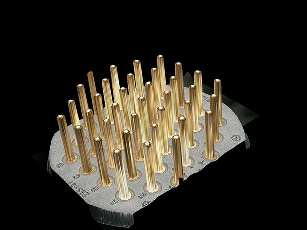
Non-destructive observation is possible; there is no need to cut or put a sample on a slide to prepare it for observation. The VHX Series Digital Microscope naturally has a 20x larger depth of field compared to conventional optical microscopes, so users can view a part like a 9-pin connector fully in focus at any angle. At high magnifications, users are still able to capture a fully-focused image despite the naturally smaller depth of field due to the depth composition function.
Tilted observation of threads

Macro image, 20x

Magnified image, 200x

Larger Working Distance Allows for Free-Angle Observation
With the VHX Series Digital Microscope, users are able to view a sample from any angle! You no longer need to create a fixture or use putty to observe your sample from the most optical angle - just tilt the optics.
Same Quality - Better Observation Experience
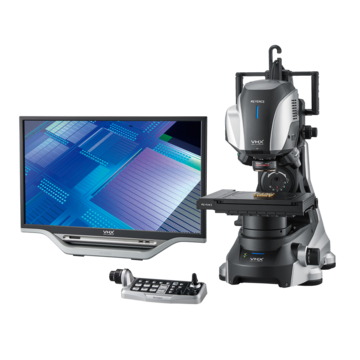
Unlike traditional optical microscopes, the VHX Series Digital Microscope uses a camera and lens to display the sample on a large monitor. This allows for easier collaboration with lab members while still maintaining the same image quality.


Accurate Measurements With Automated Reporting
With the VHX Series Digital Microscope, users can easily capture 2D/3D measurements that can be automatically exported and input to a report.
Measurement of the graphite spheroidization rate, 100x


2D measurement image
3D profile of connector pins, 50x

3D measurement image