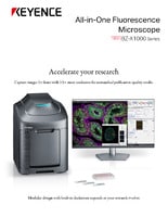
Fluorescence Microscopes
Stitching, Sectioning and the Z-Stack Function as Decisive Arguments for the Acquisition of the BZ Fluorescence Microscope at the University Hospital of Düsseldorf

Prof. Dr. Norbert Gattermann
Managing Director of the University Cancer Center (UTZ)
Senior Physician at the Department of Hematology, Oncology and Clinical Immunology
University Hospital of Düsseldorf
Professor Norbert Gattermann is the Managing Director of the University Cancer Center (UTZ) in Düsseldorf. His research focuses on malignant diseases in bone marrow, primarily preleukemias, also known as myelodysplastic syndromes (MDS). Since a certain form of MDS in bone marrow is characterized by pronounced iron overload in the mitochondria of erythroblasts (maturing progenitor cells of red blood cells), he is also interested in specific issues relating to iron metabolism.
For more than 30 years, the University Hospital of Düsseldorf has been known for its research on MDS. It is home to a large MDS registry, which focuses on careful long-term documentation of the course of the diseases and provides an excellent basis for research on the epidemiology, diagnostics and risk assessment of these illnesses. A large number of bone marrow and blood samples have been collected and stored in the MDS registry’s biomaterial database by Norbert Gattermann and his colleagues over many years, and they can now be used for molecular biological analysis.
Get detailed information on our products by downloading our catalog.
View Catalog

Tissue microarray of bone marrow punch biopsies and 3D cell cultures are a particular challenge in microscopy
The MDS working group consists of 2 Masters students, a Physician-Scientist and a Biological Technical Assistant (BTA).
Gattermann says: “The BZ fluorescence microscope impressed us not only because of its value for money, but also because it can be expanded with different modules. We’re a small working group, and we’re very grateful that we were able to get funds from a foundation to purchase it. Little by little, we’ll be able to integrate new modules.”
The members of the hemato-oncology group conduct research at the cellular level on three foci, so an upright fluorescence microscope is already part of their standard equipment, and they successfully carry out Opal multiplex IHC assays with up to seven fluorescent dyes. But this microscope does not meet all their requirements, which was one of the key arguments for acquiring the BZ fluorescence microscope.
One distinctive result of the research group’s work is the specially developed tissue microarrays (TMAs) of MDS bone marrow punch biopsies, which contain biopsies from patients with AML (acute myeloid leukemia) and normal controls for direct comparison. When evaluating immunohistochemical studies on the TMAs, one major advantage of the BZ fluorescence microscope is that one – or even multiple – samples on the TMA can be captured together in one image.

Figure 1: TMA stitch DAPI + R-loop, edited

Figure 2: TMA section stitch DAPI + R-loop, edited

Figure 3: TMA spots overview
“The stitching function is particularly important for us in order to be able to display the 2 mm core in an image,” explains Gattermann.
Stitching is also useful for evaluating in vitro cultures in bone marrow progenitor cells, where for example we study the mitochondria of erythroblasts as the target structure. A major advantage of the BZ fluorescence microscope for this type of preparation is of course the microscope’s inverted beam path, which allows easy positioning and evaluation of the cell cultures. And Simon Brille (BTA) adds: “The microscope and software are very intuitive to use. The analysis software gives us various ways to process images quickly and easily.”
We’re here to provide you with more details.
Reach out today!

1. Pathophysiology of sideroblastic anemia
Sideroblastic anemia is a special form of MDS which involves a massive iron overload of the mitochondria in the erythropoiesis progenitor cells. Although this disorder has been known since the 1950s, the mechanics of it are still unclear. Several years ago, spliceosome mutations in the SF3B1 gene were found to be present in 75-80% of all patients with sideroblastic anemia. This means that although the mitochondria absorb iron, the enzyme ferrochelatase does not incorporate the iron into protoporphyrin as effectively as it should and the iron accumulates in the mitochondrial matrix. As a result, far too little heme synthesis takes place. Without heme, the cell signals that iron is still missing, leading to further flooding of the mitochondria with iron. The excess iron oxidizes and damages the mitochondria.
Improving our understanding of interactions at the mitochondrial membrane
Gattermann and his fellow scientists are conducting research on protein interactions at the inner mitochondrial membrane and in the matrix to find out why the iron and porphyrin do not combine properly. They use the BZ fluorescence microscope to examine membrane complexes for structural abnormalities by visualizing protein-protein interactions as fluorescent signals with the proximity ligation assay (PLA). The sectioning function produces clear images without fluorescence blurring. Only the signals from the focused planes are extracted and used to create a fully focused image. The protein interactions studied involve ferrochelatase and its interaction partners in the “mitochondrial heme metabolism complex”, some of which are only temporary.

Figure 4: Proximity ligation assay
“While visiting a working group in Bonn, we stumbled across a presentation of the BZ fluorescence microscope and were immediately fascinated by everything this compact device has to offer!” recalls Fatemeh Majidi, Physician Scientist.
We’re here to provide you with more details.
Reach out today!

2. Pathophysiology in myelodysplastic syndrome in the 3D cell culture
In order to realistically represent and understand the behavior of bone marrow progenitor cells, Gattermann and his working group are currently establishing the 3D cell culture technology in his laboratory. Previously, the usual two-dimensional colony forming assay was carried out, a method in which hematopoietic progenitor cells form colonies in a semi-solid medium and can be studied in that state. The 3D cell culture has further increased the demands on microscopy, and it's where the stitching, sectioning, and Z-stack functions of the BZ fluorescence microscope really show their value. These functions were decisive arguments for the acquisition of the BZ fluorescence microscope. Gattermann and his colleagues are opening up brand new methods of visualization for applications in immunohistochemistry, nuclear staining, and detecting DNA-RNA hybrid structures in the 3D cell culture.
We’re here to provide you with more details.
Reach out today!

3. Drug development: neddylation inhibitor
A neddylation inhibitor, the effect of which has not yet been fully explored, is currently being tested in clinical trials. Gattermann’s working group is also investigating how this new drug affects hematopoietic progenitor cells and their differentiation. Among other things, the proximity ligation assay (PLA) on the epidermal growth factor receptor (EGFR) detects homodimerization of the receptor on the cell surface, which results in receptor phosphorylation in the cell interior and therefore has a signaling effect in terms of cell proliferation. At the time of the interview with Professor Gattermann, it was not yet possible to provide more precise information because the initial results were still in the process of being published.
The acquisition of the BZ fluorescence microscope is a major step forward for Norbert Gattermann’s working group. Translational projects within the University of Düsseldorf will ensure other researchers can use the BZ fluorescence microscope.


