Fluorescence Microscopes
Quantifying Hyphal Concentration Using an Image Cytometer (ICM)
-
Tags:
- Microbiology , Food
What are image cytometers?
There are two major categories of cytometry: flow cytometry (FCM) and image cytometry (ICM). Instruments that use these measurement methods are called flow cytometers and image cytometers, respectively.
Image cytometers are systems that can quickly quantify large populations of cells or particulates by capturing and analyzing images. Unlike flow cytometry, cells can be kept in whatever media they are typically contained (slides, dishes, plates, etc.) - there is no need for them to be removed. The position of each cell on a well plate or slide and the measurement information are recorded, allowing users to quantify and visually compare results.
Get detailed information on our products by downloading our catalog.
View Catalog

Limitations of flow cytometers
Flow cytometers are used at many laboratories due to their ability to quickly count a large number of cells. However, these systems are not without issues, including: 1) inability to visually confirm measurement results and 2) cells must be removed from their natural state in order to be analyzed. Additionally, flow cytometers can be clogged when evaluating larger cells or those that have adhesive properties.

Advantages of image cytometers
As imaging technology has improved over the past several years, image cytometers have been receiving more attention. Unlike flow cytometers, image cytometers are able to capture an actual image of the sample, quantify, and analyze cellular phenomena without any need to remove cultured cells from their containers. These systems enable quantification in conjunction with imaging, such as linking and analyzing a cell's shape and the localization and manifestation information of multiple proteins, which was conventionally very difficult to perform.
Automated measurement with an image cytometer
When attempting to quantify cells on a well plate or slide, conventional cytometers could not produce accurate measurements, were prone to user variation, or could not capture an image of the entire sample.
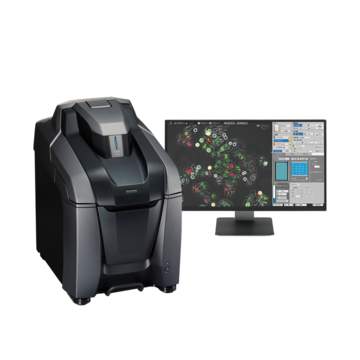
The BZ-X800 is equipped with the latest image cytometer functions, allowing the system to perform high-precision quantification and imaging similar to a confocal microscope for improved analysis.
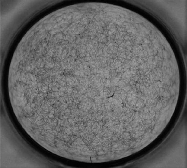
Magnified image of hyphae

96-well plate
Quantification result display
The parts indicated in red on the heat map show the wells with a high concentration of hyphae

Hyphal density is quantified by transmittance (brightness).
Wells with low brightness have a high density of hyphae.
Using the All-in-One Fluorescence Microscope BZ-X800
- The BZ-X800 enables accurate quantification as well as analysis of hyphal growth and concentration.
- An entire well can be captured at high-magnification, enabling the user to quickly see cellular distribution and characteristics.
- With fully-motorized operation and an integrated darkroom, anyone can easily operate the instrument to produce advanced analysis with no user-dependent variations.